
Biological Research Using SWIR Camera Technology
NIR-SWIR wavelengths have become important for biological research and clinical studies. Whether it is because of reduced light scattering at longer wavelengths, for detecting molecular (chemical) absorption bands or for capturing the emissions of multiple fluorescent dyes. Reduced scatter permits the light probes of Optical Coherence Tomography (OCT) to penetrate deeper in otherwise opaque tissues for imaging the layers of the retina. The molecular absorption band of water permits imaging through the opaque eardrum or through the top layer of skin to see the buildup of fluid which is causing earaches or causing wheals or hives on the skin, thus marking an allergic reaction. In another application, SWIR's reduced scatter and ability to capture infrared fluorescent dye emissions enables imagine-guided surgery, even when a tumor is under a layer of healthy tissue. NIR and SWIR fluorescence dyes are used in this technique to tag different tissues, helping the surgeon identify and avoid damaging nerves, bile ducts, blood vessels or other organs while still identifying all the cancerous tumor.
By using a filter wheel, or even by depositing filters in a pattern on the detector’s pixels, the wide visible to SWIR wavelength range of our SWIR cameras can be employed to permit one optical path and sensor to image several visible, NIR or SWIR dyes at once. The SWIR camera then accurately captures the position of each tagged tissue and avoids the risk of mechanical misalignments or miscalibrations, which could mislead the surgeon. The wide dynamic range of our SWIR cameras and their 14-bit linearity permit imaging complex surgical fields with the image quality to notice the weakest glows in the same field of view as a bright reflection of the operating theater lamps. Megapixel formats with sensitive 12 µm pixels give the resolution and field of view needed for accurate surgery.
Our 1280SciCam scientific SWIR camera offers cooled operation when dealing with very weak emissions while the MVCam SWIR camera is small and compact, for fitting into the limited space over the patient. The sensor chip itself can be used for multi-spectral or hyperspectral imaging with the addition of appropriate wavelength-selective optics (call our applications group to discuss custom solutions for your application). With a simple bandpass filter for the water band, the MVCam SWIR camera can be implemented in an allergist office for imaging the allergy induced wheals or bumps to provide a digital record of the patient’s reaction.
An important biological use of InGaAs photodiode linear arrays is for Spectral Domain Optical Coherence Tomography (SD-OCT). Here coherent light from a fiber optic light source is used to image beneath scattering layers of tissue or materials. InGaAs photodiodes permit longer wavelengths to be used, which better penetrate tissues and enable imaging all the layers of the retina or even measure the distance from the cornea to the back of the eye. In this technique the light backscattering from structures in the tissue is collected by the same fiber and filtered by interferometer, then passed through a spectrometer onto a linear photodiode array. This generates a depth profile scan of the tissue structure after a mathematical process called a Fast Fourier Transform (FFT) decodes the spectral interferogram. Then by scanning the laser spot across the tissue, a 3-D image is constructed that can be examined by the eye doctor in ways like a CAT scan or MRI.

 MVCam
MVCam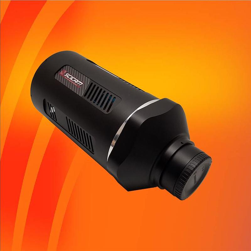 1280SciCam